Enhanced Visual System of Butterflies Inspires New Bioinspired Imaging Sensor
Published in the journal Science Advances, a recent study led by Professor Viktor Gruev and Professor Shuming Nie from the University of Illinois Urbana-Champaign introduces a groundbreaking imaging sensor that replicates the enhanced visual system of butterflies. This sensor is capable of “seeing” into the ultraviolet (UV) range, which is inaccessible to human eyes.
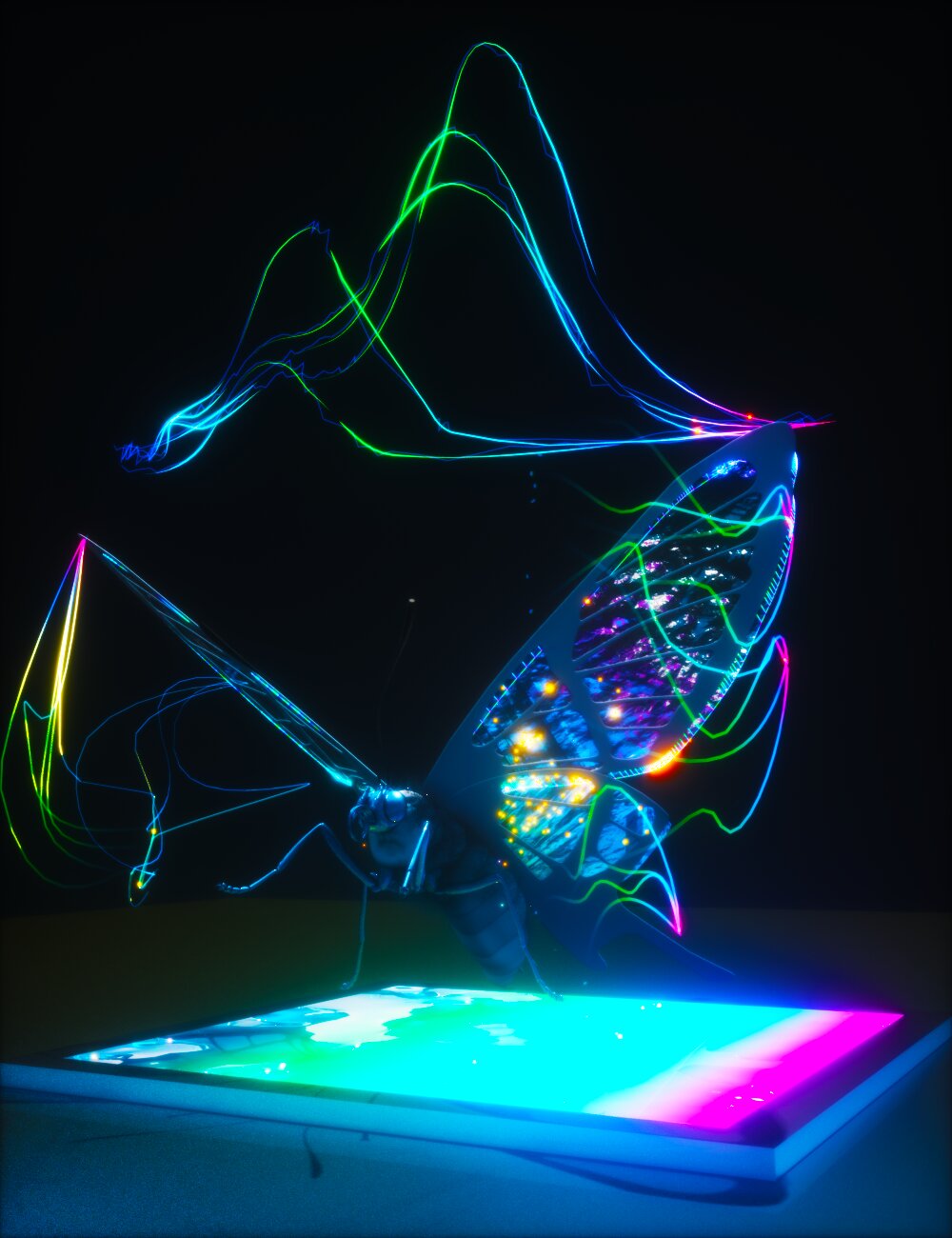
Butterflies possess a visual system that allows them to perceive a broader range of colors, including UV light. Taking inspiration from this, the research team designed a camera using stacked photodiodes and perovskite nanocrystals (PNCs) that can detect multiple UV regions. PNCs are semiconductor nanocrystals that exhibit unique properties for sensing applications.
Whereas humans have trichromatic vision with three photoreceptors, butterflies have compound eyes with six or more photoreceptor classes that have distinct spectral sensitivities. The Papilio xuthus butterfly, for example, has blue, green, red, violet, ultraviolet, and broadband receptors. Additionally, butterflies have fluorescent pigments that convert UV light into visible light, enabling them to perceive a wider range of colors.
The imaging sensor developed by the UIUC team emulates the UV sensing mechanism of the Papilio xuthus butterfly by combining a layer of PNCs with a tiered array of silicon photodiodes. The PNC layer absorbs UV photons and emits visible light, which is then detected by the tiered silicon photodiodes. Through signal processing, UV signatures can be mapped and identified.
This new technology has significant implications in the field of healthcare. By exciting certain biomedical markers, such as amino acids, proteins, and enzymes, with UV light, these markers fluoresce and emit UV and visible light in a process called autofluorescence. Cancerous cells typically have higher concentrations of these markers compared to healthy cells, resulting in different spectral signatures. The imaging sensor can differentiate between cancer and healthy cells with 99% confidence.
The UIUC team envisions the use of this sensor during surgery to aid in the decision-making process of removing cancerous tumors. It can help surgeons determine the appropriate amount of tissue to remove for clear margins. Furthermore, this technology opens up opportunities in fields beyond healthcare, such as biology and underwater exploration. Understanding how other species use UV light and detecting UV underwater can provide valuable insights.
This bioinspired imaging sensor represents a significant technological advancement, allowing humans to perceive the world in ways previously inaccessible.
More information:
Cheng Chen et al, Bioinspired, vertically stacked, and perovskite nanocrystal–enhanced CMOS imaging sensors for resolving UV spectral signatures, Science Advances (2023). DOI: 10.1126/sciadv.adk3860. www.science.org/doi/10.1126/sciadv.adk3860
